European Society for Magnetic Resonance in Medicine and Biology

PRECONGRESS WORKSHOPS
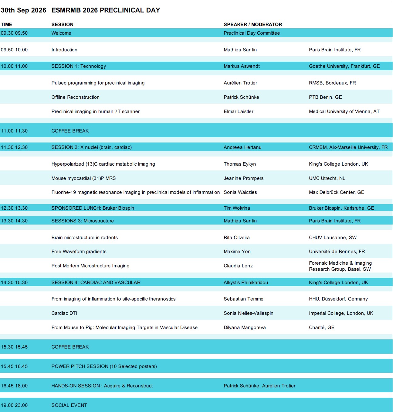
Preclinical MRI Frontiers: Shaping the Future Together
Full day workshop
Chair: Mathieu D . Santin (FR)
Committee: Andreea Hertanu (FR), Markus Aswendt (DE), Alkystis Phinikaridou (UK)
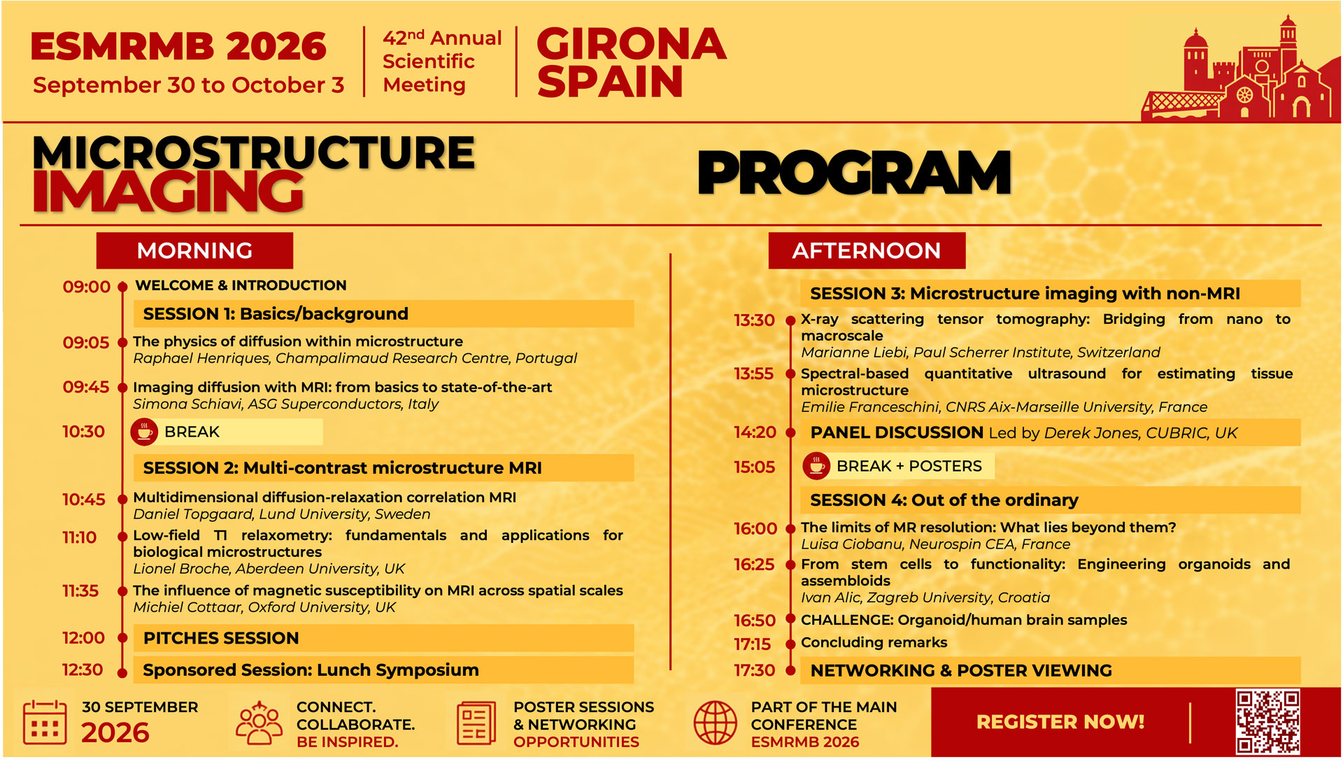
Microstructure imaging
Full day workshop
Chairs: Chantal Tax (NL & UK), Mariam Andersson (DK)
Committee: Elda Fischi-Gómez (CH), Emma Thomson (DK), Maëliss Jallais (UK), Isabelle Corouge (FR)
This whole-day workshop will provide a structured overview of state-of-the-art microstructure imaging, integrating physical foundations, methodological innovation, and complementary validation strategies, aiming to support interaction between communities that may otherwise operate separately. The target audience includes researchers, clinician-scientists, engineers, and trainees working in or entering the field of microstructure imaging.
Microstructure imaging is among the most rapidly advancing domains in magnetic resonance research, enabling increasingly specific insights into tissue architecture and pathological alterations. Despite these advances, important challenges persist regarding model assumptions, biological specificity, and cross-modal validation.
The workshop will open with an interactive session on the physics of diffusion in microstructure, introducing propagator formalism and diffusion encoding strategies. The second session will focus on multi-contrast microstructure MRI, including diffusion–relaxation methods, low-field relaxometry, and diffusion-QSM, emphasising how integrated contrasts enhance sensitivity to specific microstructural features. The third session will broaden the perspective of microstructure imaging beyond MRI, incorporating talks on advanced microscopy and ultrasound approaches by leading researchers within their respective fields, followed by a panel discussion. The final session will highlight emerging directions, including within-cell MRI and organoid-based microstructure imaging, and will introduce a proposed community-wide microstructure imaging challenge designed to stimulate methodological benchmarking and development, as well as collaborative discussion.
Abstract presenters will be invited to deliver short pitches, complemented by dedicated poster-viewing sessions.
Microstructure workshop posters
#53688 An open-source tool creating realistic digital twins of cells for diffusion MRI modeling. Malte Brammerloh, Department of Radiology, Lausanne University Hospital (CHUV), Lausanne, Switzerland
#54115 Accounting for susceptibility effects in chemical-shift encoded fat-water separation for muscular dystrophies. Pierre-Yves Baudin, Institute of Myology, Paris, France
#54138 k-q-ASeDiWA: Joint k-q reconstruction for accelerated diffusion MRI with learned phase estimation. Anni Liu, Bruker BioSpin GmbH & Co. KG, Ettlingen, Germany, Bio-Imaging
#54208 To tilt or not to tilt: Assessing the impact of physical head misalignment on orientation-dependence of MRI parameters. Melanie BAUER, Department of Radiology, Medical University of Innsbruck, Innsbruck, Austria
#54274 Joint diffusion-relaxometry of the brain via multi-echo spin-echo diffusion EPI. Aron Gimesi, Instituto Superior Técnico, Lisboa, Portugal
#54308 Distinct microstructural diffusion MRI signatures in directed and undirected cortical organoids. Andrés le Boeuf Flo, Centre for Biomedical Imaging, SP CHUV-EPFL Section, Lausanne, Switzerland, Department of Radiology, University Hospital and University of Lausanne, Switzerland, Signal Processing Laboratory 5 (LTS5), École Polytechnique Fédérale de Lausanne, Switzerland
#54323 Reassessing intracellular volume fractions by combining diffusion MRI with relaxation time component analysis. Bastian Maus, University of Münster, Clinic of Radiology, Experimental Magnetic Resonance, Münster, Germany
#54339 μNN: a physics-informed histology-MR neural network for microstructural diffusion imaging in cancer. Athanasios Grigoriou, Institute of Oncology (VHIO), Hospital Campus, Instituto de Investigación Sanitaria Hospital Universitari Vall d’Hebron (IIS IR-HUVH), University of Barcelona, Barcelona, Spain, Center for Biomedical Imaging CIBM SP CHUV-EPFL Section, Lausanne, Switzerland
#54341 Human-informed white matter microstructural simulations link myelin loss to diffusion metrics. Alessandro Di Matteo, Biomedical Physics, Amsterdam UMC, Amsterdam, The Netherlands
#54355 Mapping liver microstructure with Histo-µSim: Effect of regularization and maximum b-value. Matthew Cherukara, King’s College London, London, United Kingdom
#54362 Deep Learning Synthesis of Diffusion Gradient Directions for Accelerated Diffusion Tensor Imaging. Simone Santoro, Department of Computer Science, University of Pisa, Pisa, Italy, Italian National Institute for Nuclear Physics, Section of Bologna, Bologna, Italy
#54381 Non-invasive glioma grading through biophysical diffusion MRI modelling:the complementary role of Histo-?Sim and SANDI. Athanasios Grigoriou, Radiomics Group, Institute of Oncology (VHIO), Hospital Campus, Vall d’Hebron Barcelona, Center for Biomedical Imaging CIBM SP CHUV-EPFL, Lausanne, Switzerland, Department of Biomedicine
#54401 Contrast ratio assessment of the Substantia Niga in Neuromelanin-Sensitive MRI using TSE and MT-GRE sequences. Vahid Malekian, IXICO plc, London, United Kingdom
#54423 Advanced Diffusion Parameter Mapping Using a Mobile 1.5T MRI Scanner. Christoph S Aigner, MPI for Human Development, Physikalisch-Technische Bundesanstalt (PTB), Berlin, Germany
#54452 Parameter-Estimation-Driven Optimization of Diffusion MRI Protocols for Microstructure Imaging Ekin Taskin, Signal Processing Laboratory 5 (LTS5), École Polytechnique Fédérale de Lausanne, Lausanne, Switzerland
#54457 Functional and structural brain correlates in obesity: Multimodal neuroimaging reveals insulo-striatal reward hyperconnectivity and disruption of memory-interoceptive pathways associated with microstructure and magnetic susceptibility. Elena de la Calle, Department of Radiology, Hospital Universitari de Girona Doctor Josep Trueta, Institut de Diagnòstic per la Imatge (IDI), Girona, Spain, Medical Imaging Research Group, Girona Biomedical Research Institute (IDIBGI), Salt, Spain
#54459 Altered association between grey matter microstructural characteristics and cerebral oxidative metabolism in Multiple Sclerosis. Alessandra S Caporale, Department of Neuroscience, Imaging and Clinical Sciences, Institute for Advanced Biomedical Technologies (ITAB), ?G.d’Annunzio’ University of Chieti-Pescara, Chieti, Italy
#54464 Fluid-suppressed QTI unmasks isotropic diffusivity variance in high-grade glioma: initial results. Oliver Gödicke, Division of Medical Physics in Radiology, German Cancer Research Center (DKFZ), Faculty of Physics and Astronomy, Heidelberg University, Heidelberg, Germany
#54492 Simulating how tissue microstructure affects multimodal MRI. Michiel Cottaar, Oxford Centre for Integrative Neuroimaging, University of Oxford, Oxford, United Kingdom
#54503 Revealing the myelin-induced magnetic field correlation modulated by white matter tract orientation at ultra-high field. Lewis Kitchingman, Cardiff University, Cardiff, United Kingdom
#54519 Neuroinflammation-related microstructural alterations using kurtosis time-dependence. Paulina J Villaseñor, Division of Neuroscience, University of Manchester, Manchester, United Kingdom
#54521 Exact PGSE Signal Model for Diffusion Confined to Cylindrical Surfaces. Erick Jorge Canales-Rodríguez, Department of Signal Theory, Networking and Communications, University of Granada, Granada, Spain
#54534 Co-MEDS: Convex Maximum Entropy Deconvolution on a Sphere for Fiber Orientation Estimation in Diffusion MRI. Harshit Harshit, Institut de mathématiques de Toulouse, Univ Toulouse, CNRS, CerCo, Toulouse, France
#54540 In vivo detection of extracellular microscopic tortuosity in the human corpus callosum using oscillating-gradient diffusion filters at 3T. Ignacio Lembo Ferrari, Center for Mind/Brain Sciences – CIMeC, University of Trento, Rovereto, Italy, Bariloche Atomic Center, CONICET, CNEA, Bariloche, Argentina
#54549 Coronary artery revascularization type and disease severity is linked with alterations in normal-appearing white matter microstructure. Ali Rezaei, School of Health, Concordia University, Centre Epic and Research Center, Montreal Heart Institute, Montreal, Canada
#54565 Probabilistic DL approaches in Diffusion-Relaxation MRI model fitting: Comparison Of Mixture Density Networks and Normalizing Flows. Nicola Casali, Politecnico di Milano; STIIMA Consiglio Nazionale delle Ricerche, Milan, Italy
#54588 In vivo SANDI diffusion MRI reveals cerebral neuroplasticity associated with long-term functional recovery in a rat model of spinal cord injury. Lori Berckmans, Bio-Imaging Lab, µNEURO Center for Excellence, University of Antwerp, Antwerp, Belgium
#54595 Characterizating spatiotemporal drift in Mean Diffusivity in Human and Phantom data. Manfredi Alberti, Max Planck Institute for Biological Cybernetics, Tübingen, Germany
#54597 Towards diffusion-weighted microstructure characterization of lymph nodes using simulation-based dictionary matching. Salomé Baup, Signal Processing Laboratory (LTS5), Ecole Polytechnique Fédérale de Lausanne (EPFL), Lausanne, Switzerland
#54607 Magnetization transfer imaging reveals distinct temporal microstructural changes in a rat model of intracerebral hemorrhage. Gemma Manich, Department of Morphological Sciences, Autonomous University of Barcelona, Cerdanyola del Vallès, Spain
#54613 Estimation of SV2A PET-Derived Synaptic Density from Quantitative MRI Using Data-Driven Methods. Mikhail Zubkov, GIGA-Neuroscience, CRC-Human Imaging, University of Liège, Liège / Luik, Belgium
#54624 Surface-to-Volume Ratio Mapping of Brain Organoids and Fiber Phantoms Using 28.2T High-Frequency OGSE. Tatiana Nikolaeva, Center for Image Sciences, University Medical Center Utrecht, Utrecht, The Netherlands
#54633 A numerical simulation framework for combined diffusion and perfusion MRI. Jasmine Nguyen-Duc, CHUV, Lausanne, Switzerland
#54645 Magnetic Resonance Imaging of Early Microstructural Changes in Amyotrophic Lateral Sclerosis. Nayereh Ghazi, Center for Functionally Integrative Neuroscience, Department of Clinical Medicine, Aarhus University, Aarhus, Denmark
#54646 Reliability Framework for Simulation-Based Microstructure Estimation with Real Data. Jonathan Rafael-Patino, Signal Processing Laboratory (LTS5), Ecole Polytechnique Fédérale de Lausanne (EPFL), Lausanne, Switzerland
#54681 Combined estimation of T2 and T2* using a GESSE sequence and EPG signal modelling. Quentin Raynaud, Laboratory for Research in Neuroimaging, Department for Clinical Neuroscience, Lausanne University Hospital and University of Lausanne, Lausanne, Switzerland
#54682 How does the menstrual cycle reshape women’s brains? Impact of hormonal changes on white matter microstructure with free water elimination. Rita Reis Nunes, Institute for Systems and Robotics and Department of Bioengineering, Instituto Superior Técnico, Universidade de Lisboa, Lisboa, Portugal
#54686 Under one minute NATIve QSM enables robust and reproducible subcortical iron mapping with strong clinical translation potential. Jazmin Ximena Suarez Revelo, Grupo de Investigación en Imágenes e IA en Salud – SURA, Ayudas diagnósticas , Medellin, Colombia
#54693 Evaluating MTV-correction in MRI models for in vivo brain iron estimation. Maryana Pozina, Edmond and Lily Safra Center for Brain Sciences, The Hebrew University, Jerusalem, Israel
#54694 Decoding the female brain: Investigating cerebral white matter changes throughout the menopausal transition, using diffusion MRI. Kato Herregods, Dep. Diagnostic Sciences, Ghent, Belgium
#54701 GAIA – Green Artificial Intelligence for Accelerated medical imaging: Sustainable and efficient diffusion MRI analysis. Maeliss Jallais, Cardiff University, Cardiff, United Kingdom
#54710 Effect of Effective Frequency Definition on Surface-to-Volume Ratio Estimation from Oscillating Gradient Diffusion MRI. Maria Paula Del Popolo, UMC Utrecht, Utrecht, The Netherlands
#54142 Comparison of Diagnostic Performance of Malignant from Benign Prostatic Areas on Prostatic DWIs with Different b-Values between Complexed Signal Average and Conventional Reconstruction Methods. Yoshiharu Ohno, Fujita Health University School of Medicine, Toyoake, Japan
#54248 Differential vulnerability of ODF- and streamline-based tractography metrics to noise levels in diffusion MRI. Sewon Lim, Department of Health Science, General Graduate School of Gachon university, Incheon, Republic of Korea
#54256 Impact of moderate Multiband Acceleration on Quality of Diffusion MRI Quality of the Brain at 3T. Marco Meixner, Science Hub Neuroscience, Ruhr University Bochum, St. Josef Hospital, Institute of Neuroradiology, Bochum, Germany
#54286 Evaluation of IVIM parameter recoverability using limited four-b-value diffusion MRI in esophageal cancer. Victoria Joppin, Department of Radiodiagnostic and Interventional Radiology, Lausanne University Hospital and University of Lausanne, Lausanne, Switzerland
#54329 Ex vivo microstructural characterization of oral squamous cell carcinoma using IMPULSED time-dependent diffusion MRI. Jeroen de Groot, University Medical Center Utrecht, Utrecht, The Netherlands
#54425 Lithium-associated white matter microstructural changes in bipolar disorder: preliminary TBSS-tractometry results from the R-LiNk study. Giovanni Videtta, Department of Neurosciences and Mental Health, Fondazione IRCCS Ca’ Granda Ospedale Maggiore Policlinico, Milan, Italy
#54512 Impact of regularized parallel imaging reconstruction on cardiac diffusion tensor imaging parameter maps. Vitali Telezki, Institute for Cardiovascular Imaging, Center of Clinical Radiology Imaging, University Medical Center Göttingen, German Centre for Cardiovascular Research (DZHK), partner site Lower Saxony, Göttingen, Germany
#54523 White-matter brain-age gap from along-tract FA and MD and its association with cognitive development in youth. Sophie Mosegaard, LTS5, École Polytechnique Fédérale de Lausanne (EPFL), Lausanne, Switzerland
#54571 Preliminary Evaluation of Fractional Anisotropy Sensitivity to Longitudinal Changes Following Peripheral Nerve Injury. Isaac Manzanera Esteve, Plastic Surgery, Vanderbilt University Medical Center, Nashville, USA
#54584 Detectability analysis of lymphocyte infiltration measurement via clinical diffusion MRI: a simulation study. Daniel Navarro-Garcia, Vall d’Hebron Institute of Oncology, Barcelona, Spain
#54593 Structural Connectome Sensitivity Analysis in the Alzheimer’s Disease Spectrum using Cortical ROI Amendment for Juxtacortical White Matter Parcellation. Gyula Gyebnár, Semmelweis University Medical Imaging Centre, Budapest, Hungary
#54649 Neonatal white matter FA in thalamo-frontal and perirolandic tracts predicts full-scale IQ at age 5 in children with spina bifida aperta. Tobias Fernandez-Borkel, Center for MR Research, University Children’s Hospital Zurich, Neuroscience Center Zurich, Zurich, Switzerland
#54140 How To Improve Prediction Capability for Patient’s Outcome in Postoperative Stage I NSCLC Patients with Different Molecular Information on MRI with PET/CT. Masahiro Endo, Shizuoka Cancer Center, Sunto-gun, Nagaizumi-cho, Japan
#54213 Early detection of bone metastases in prostate cancer using advanced whole-body dMRI. Carlos Macarro, Vall d’Hebron Institute of Oncology, University of Barcelona, Barcelona, Spain
#54356 Incomplete reversibility of fixation-induced changes in postmortem human brain white matter MRI following rehydration. Lennart Bedarf, Institute of Forensic Medicine, Department of Biomedical Engineering, University of Basel, Institute of Forensic Medicine, Health Department Basel-Stadt, Basel, Switzerland
#54406 Spatial-angular implicit neural representations for accelerated diffusion MRI via joint k-q reconstruction. Lara Kunze, imec-Vision Lab, Department of Physics, University of Antwerp, Antwerpen, Belgium
#54605 Multiparametric characterization of cervical spinal cord involvement in women with X-linked adrenoleukodystrophy: preliminary results from the ARISE study. Marianne Golse, Neuroradiology department, Hôpital de la Pitié-Salpétrière, Laboratoire d’Imagerie Biomédicale, Sorbonne University, Paris, France
#55453 A unified preprocessing framework for preclinical microstructural and quantitative MRI data. Cristiana Tisca, Oxford Centre for Integrative Neuroimaging, FMRIB, Nuffield Department of Clinical Neurosciences, University of Oxford, Oxford, Royaume-Uni
#55474 DEMONE: A longitudinal mouse-to-human study linking brain imaging biomarkers to cellular pathology in Myotonic Dystrophy Type 1. Eléonore Vermeulen, Centre de NeuroImagerie de Recherche – CENIR, Paris Brain Institute – ICM, Paris, France
Practical MRI Safety Management
Organizers
Allison McGee,
University College Dublin
allison.mcgee@ucd.ie
Claude Portanier,
Mater Dei Hospital, Malta
claude.portanier-mifsud@gov.mt
Duration
Half day in the morning from 09:00 to 12:30
Target audience
This session will be of interest to the MRI community, including researchers, medical physicists, clinicians and radiographers.
Session Schedule
Please see below the proposed schedule with speaker names for those who have agreed to contribute if this pre-congress session is approved by the CPC.
09:00 Workshop Introduction (Allison McGee, Ireland)
09:10 ESMRMB practice guidelines when managing MR safety in vulnerable patients (Francesco Santini, Basel)
09:40 Safety considerations for remote MRI (Tomasz Bienias, Germany)
10:10 Safety considerations in paediatric MRI (Fernaiza Sahidjuan, GOSH London)
10:40 Q&A session 1
10:55 Coffee Break
11:15 MR scanning protocol optimisation for MR conditional implants (Claude Portanier Mifsud, Malta)
11:45 Considerations and processes for hearing implant MRI testing (Markus Nagl, Med-EL Medical Electronics Austria)
12:15 Q&A session 2
12:30 Close
MRI in Multiple Sclerosis and Related Neuroimmunological Disorders: Translating Clinical Needs into Robust MR Solutions
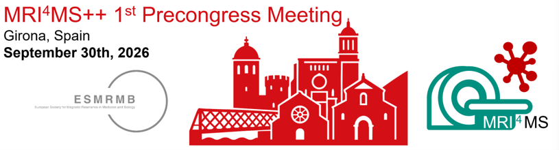
Half-day workshop
Chair: Sonia Waiczies (DE)
Committee: Ismail Koubiyr (NL), Myrte Strik (NL)
Annual Meeting of the Working Group on Multiple Sclerosis and Related Neuroimmunological Disorders: Translating Clinical Needs into Robust MR Solutions
This meeting focuses on how advanced MRI can support diagnosis, prognostics, and therapy monitoring in MS, with translational extension to related autoimmune CNS diseases such as NMOSD and MOGAD. We aim to bring together MR physicists, imaging scientists, neurologists, neuroradiologists, and industry stakeholders, fostering exchange between clinical needs, biomarker development, and implementation.
| Session 1: Optic Nerve | 13:30 – 14:15 |
Olivier Outteryck, Université de Lille, FR Bringing Optic Nerve MRI to the Foreground: a Clinical Perspective Deborah Pareto, Vall d’Hebron University Hospital, ES Bringing the Optic Nerve into Focus: Technical and Quantitative MRI in MS | |
| Session 2: MR Lesion Biomarkers | 14:15 – 15:00 |
Alessandro Cagol, University of Basel, CH Paramagnetic Rim Lesions and Slowly Expanding Lesions in MS: from MRI Biomarkers to Clinical Impact Meritxell Bach Cuadra, Lausanne University, CH Paramagnetic Rim Lesions and Slowly Expanding Lesions in MS: from MRI Acquisition | |
| Power pitch presentations of selected abstracts | 15:00 – 15:30 |
Emilie Poiron, Rothschild Foundation Hospital, FR Elise Bannier, Centre Hospitalier Universitaire de Rennes, FR Carla Xena Bosch, Universitat Oberta de Catalunya, ES Ariadna Masot-Llima, Multiple Sclerosis Centre of Catalonia, ES Francisco José Aparicio Serrano, Multiple Sclerosis Centre of Catalonia, ES | |
| Coffee Break | 15:30 – 16:00 |
| Session 3: Patient Trajectories and Prognosis | 16:00 – 16:45 |
Carmen Tur, Vall d’Hebron University Hospital, ES Understanding and Predicting Progression in MS Loredana Storelli, IRCCS San Raffaele Scientific Institute, IT Patient Trajectories and Prognosis in Neuroimmunological Disorders: A Technical Perspective on MRI-Based Modeling | |
| Session 4: MRI in Clinical and Research Settings | 16:45 – 17:30 |
Pablo Naval Baudín, Bellvitge University Hospital, ES MRI Acquisition in MS: a Clinical Perspective Frederic Sampedro, Vall d’Hebron University Hospital, ES The Evolution of MRI Acquisition: a Neuroradiologist’s Perspective | |
| Round table discussion and WG Meeting | 17:30 – 18:00 |
To participate, please register to the Pre-Conference Day Meetings here
Annual Meeting of the Glioma MRI (GliMR) Working Group
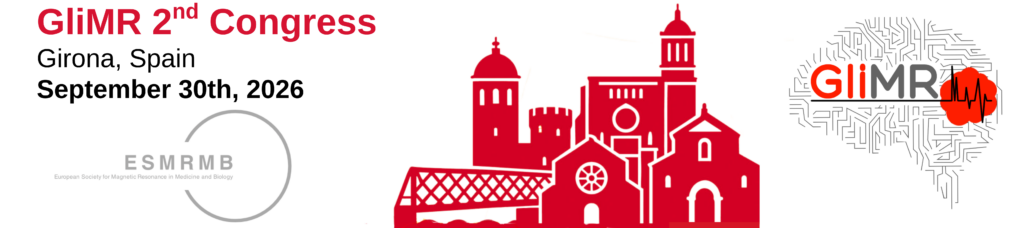
Annual Meeting of the Glioma MRI (GliMR) Working Group
The Glioma MRI community invites you to join the GliMR 2026 Pre-Day Meeting to discuss the current state and future directions of brain tumor MRI. The meeting will span clinical practice to translational neuro-oncology imaging, with a strong emphasis on trainees and early-career researchers. It will also actively engage industry partners and patient representatives, ensuring that all key stakeholders in brain tumour imaging are represented.
The event is tailored for trainees, early- and senior-career researchers, radiographers, and clinicians with a primary interest in brain tumour imaging, encompassing diverse backgrounds such as neuroimaging and data science, neuroradiology, neurosurgery, and preclinical/basic research in glioma models. The programme is structured into five sessions designed to promote knowledge exchange, highlight recent scientific and technological advances, and critically assess current challenges while identifying future directions in glioma MRI, in collaboration with industry and patient representatives.
To participate, please register for the Pre-Day Meeting: REGISTRATION
Important announcements:
- A call for networking projects (including ongoing initiatives and new proposals) is open until 11 September – extended deadline! Applications can be submitted here.
- Travel stipends are available for trainees, early-career researchers, radiographers, and clinicians (see details below). Applications must be submitted by 15 July 2026 using this application form.
Program
| Time | Sessions | Moderators / Speakers |
| 08:50-09:00 | Welcome | Esther Warnert, Rui Simoes |
| 09:00-10:30 | S1. Improve patient outcomes with brain tumor imaging | Vera Keil, Albert Pons Escoda |
| 09:00-09:20 | Treatment planning and monitoring: seeing more, seeing better | Cristina Ramos, Porto PT |
| 09:20-09:40 | Improving neurosurgery with imaging and music | Andreu Gabarros, Barcelona ES |
| 09:40-10:00 | Non-invasive imaging and patient quality of life | Simon Newman, Fleet UK and Joshua Cull, London UK |
| 10:00-10:30 | Round Table Discussion | All Session Speakers |
| 10:30-11:00 | COFFEE BREAK | |
| 11:00-12:30 | S2. Translational frontiers for brain tumor imaging | Ana P Candiota, Rui Simoes |
| 11:00-11:30 | Imaging brain tumor microstructural remodeling | Marco Palombo, Cardiff UK |
| 11:30-12:00 | AI-enhanced glioma MRI in neuroscience and neuroradiology | Esin Ozturk, Istanbul TR |
| 12:00-12:30 | Linking brain tumor imaging and histology | Peter LaViolette, Milwaukee WI USA |
| 12:30-13:30 | LUNCH | |
| 12:30-13:30 | S3. Sponsored Session: Lunch Symposium | Ana Matoso, Nuria Arias Ramos |
| 13:45-17:45 | S4 GliMR: where we stand and new challenges | Yaara Erez, Jan Petr |
| 13:45-14:00 | Present and future of GliMR | Esther Warnert, Rotterdam NL |
| 14:00-14:20 | The Current Histomolecular Classification of Gliomas: WHO 2026 and Beyond | Cristina Carrato, Badalona ES |
| 14:20-14:40 | Cancer Imaging Europe (EUCAIM): Glioblastoma | Maria Beser Robles, Valencia ES |
| 14:40-15:10 | COFFEE BREAK | |
| 15:10-16:10 | Selected abstract presentations [LIST BELOW] | (ESMRMB abstracts submitted) |
| 16:10-17:10 | Selected project presentations | (GliMR networking initiatives submitted) |
| 17:10-17:50 | S5. Task Forces | (breakout session) |
| 17:50-18:00 | Adjourn | Esther Warnert, Rui Simoes |
Abstract presentations
| Title | Presenter | Program Number |
| A neural network approach for efficient magnetic resonance vascular fingerprinting using spin- and gradient-echo dynamic susceptibility contrast MRI | Karen van der Werff | P361 |
| Taking a new angle to map microstructural collapse and occult pathology in brain tumors using orientation-dependent NODDI | Melanie Bauer | P234 |
| Two immunometabolic worlds in high-grade gliomas: interaction between immune checkpoints and tumor metabolism. | Jesús Pacheco Torres | P276 |
| Automated classification of glioma treatment response: benchmarking via modality selection and coarse tumor detection | Ana Matoso | PG183 |
| Feasibility of Tumor-Masked Structural Connectomics and Explainable Machine Learning for Assessing White Matter Disruption in Gliomas: A Pilot Study | Montalba Cristian | PG185 |
| Automated enhancing tumour extraction via T1c image subtraction: A radiomic comparison against expert delineation in post-treatment Glioma | Akashleena Chatterjee | P399 |
| Cross-modality radiomic biomarker consistency in glioma: Comparing LASSO-selected features from DCE-MRI pharmacokinetic maps and structural MRI | Akashleena Chatterjee | P398 |
| A Continuous Learning Strategy for Automated Glioblastoma Segmentation: Knowledge Transfer between Pre-Operative and Post-Operative MRI Data | Catarina Passarinho | PG186 |
| Perfusion-Discordant Hypoxia-BOLD Hotspots in the Non-Enhancing Glioblastoma Margin: Voxelwise DSC Regression and Biopsy Validation | Tristan Schmidlechner | P265 |
| Can Uncertainty Quantification Improve Trust in AI-assisted Clinical Decision-making? A Case Study in MRI-based Glioma Subtyping | Gonzalo Esteban Mosquera Rojas | P403 |
| Correlation of BBB-ASL Exchange Time with DCE-MRI Derived Permeability Parameters in Gliomas | Gülce Turhan | PG074 |
| Harnessing 3 Tesla MRI for in vivo microstructural characterization of the glioblastoma microenvironment in patient-derived mouse models | Joao F Zamith | PG169 |
| Comparison of T2 values estimated from Multi-TE M0 and Arterial Spin Labeling MRI | Ayse Irem Cetin | P334 |
Project presentations
Title | Presenter |
Reduced Use of Contrast Agents (REDUCE) | Vera Keil |
From Tissue to MRI: Imaging Biomarkers of Glioblastoma Infiltration for Precision Surgery | Pilar Sánchez Gómez |
Spatial lipid reprogramming under immunotherapy in glioblastoma | Jesús Pacheco-Torres, ES |
Openly accessible datasets for brain tumour imaging science (OADIS-BT) | Stefanie Thust |
Multimodal MRI for pediatric brain tumour diagnostics (PediTuMRI) | Yeva Prysiazhniuk |
Open Science Initiative for Perfusion Imaging (OSIPI) | Jan Petr |
Travel Stipends
1. Regulations
Travel Stipends generously supported by The Brain Tumour Charity.
Article 1 – Purpose
The GliMR 2026 Pre-Day Meeting will award ten (10) Travel Stipends to support the participation of trainees attending the meeting in Girona, Spain, on 30 September 2026.
Article 2 – Value of the Stipend
- Each Travel Stipend has a value of €150 (one hundred and fifty euros).
- The Travel Stipends are funded through the support of The Brain Tumour Charity.
- The stipend will be paid to the selected applicants following the procedures defined by the Organizing Committee, which will request awardees to provide their IBAN account details for bank transfer payment the week following the meeting.
Article 3 – Eligibility
Applicants must: (a) be students, doctoral candidates, postdoctoral researchers, residents, fellows, or other trainees; (b) have no more than eight (8) years of research or clinical experience following completion of their PhD degree (justified career breaks will be considered); and (c) submit a statement of no more than 100 words describing why they wish to attend the GliMR 2026 Pre-Day Meeting and what they expect to gain from participation. Priority will be given to first/presenting authors of submitted work (GliMR-selected ESMRMB abstracts and/or project call) to the GliMR pre-day meeting, which should be specified in the application.
Article 4 – Evaluation Criteria
Applications will be evaluated independently by the Jury according to: Relevance of the statement (0–5 points) and Expected impact of attendance on the applicant’s training and career development (0–5 points). Maximum score: 10 points.
Article 5 – Selection Procedure
- Applications will be ranked according to their total score.
- The ten (10) highest-scoring applicants will receive a Travel Stipend, conditional on attending the GliMR 2026 Pre-Day meeting.
- In the event of a tie affecting the final positions, the Jury will reach a consensus decision.
- The Jury reserves the right not to award all stipends if the quality of applications is deemed insufficient.
Article 6 – Jury
The Jury shall consist of the Organizing Committee of the GliMR 2026 Pre-Day Meeting:
Rui Simões (Porto, Portugal)
Esther Warnert (Rotterdam, Netherlands)
Ana Paula Candiota (Barcelona, Spain)
Harish Poptani (Liverpool, United Kingdom)
Yaara Erez (Ramat Gan, Israel)
Ana Matoso (Lisbon, Portugal)
Albert Pons-Escoda (Barcelona, Spain)
The Jury’s decisions are final and not subject to appeal.
Article 7 – Final Provisions
- Any matters not covered by the present Regulations shall be decided by the Jury.
- Applications must be submitted online by 15 July 2026 via this form.
- The recipients of the Travel Stipends will be announced on this webpage by 31 July 2026 and formally recognized during the GliMR 2026 Pre-Day Meeting by a member of the Jury.
- These Regulations enter into force on the date of their publication.
2. Recipients
- Catarina Passarinho
Instituto Superior Técnico, Lisbon, Portugal - Gonzalo Esteban Mosquera Rojas
Erasmus MC, Rotterdam, Netherlands - Gülce Turhan
Boğaziçi University, Istanbul, Türkiye - Harshit Harshit
Université Toulouse, Toulouse, France - João Zamith
i3S – Instituto de Investigação e Inovação em Saúde, Porto, Portugal - Karen van der Werff
Erasmus MC, Rotterdam, Netherlands - Lobke Mesdagh
Ghent University, Ghent, Belgium - Marta Padrela Loureiro
Instituto Superior Técnico, Lisbon, Portugal - Yıldız Tüzün
Boğaziçi University, Istanbul, Türkiye
3. Addendum
As only 9 of the 10 available travel stipends were awarded, the funds from the unawarded stipend (€150) have been redistributed equally among the recipients, increasing each travel stipend to €166.
From Diagnostic Imaging to Therapeutic Guidance: The Expanding Role of MRI in Radiotherapy
Half-day workshop
Chair: Lars E. Olsson (SE)
Committee: Marielle Philippens (NL), Marco Riboldi (DE), Joan Chick (UK)
Programme
13:30 – 14:00 Introduction and motivation – Why MRI in RT – Lars E. Olsson – Lund University, Lund SE
14:00 – 14:30 Differences between diagnostic MRI-physics and MRI-physics in RT-applications – Marco Riboldi, Ludwig-Maximilians-Universität, Munich DE
14:30 – 15:00 MRI in RT – workflow, coils, protocols… – Joan Chick – ICR Royal Marden, London UK
15:00 – 15:30 MRI-only RT – Hubert Gabrys – University Hospital, Zurich CH
15:30 – 16:00 Break
16:00 – 16:30 Specific MRI QA in RT – Marielle Philippens – University Medical Center, Utrecht NL
16:30 – 17:00 Functional MRI and imaging biomarkers – Petra van Houdt -NKI, Amsterdam, NL
17:00 – 17:30 MR-guided RT, MR-linac – Bas Raaymakers – University Medical Center, Utrecht NL
17:30 – 18:00 Discussion: The Expanding Role of MRI in Radiotherapy – A Panel of Speakers
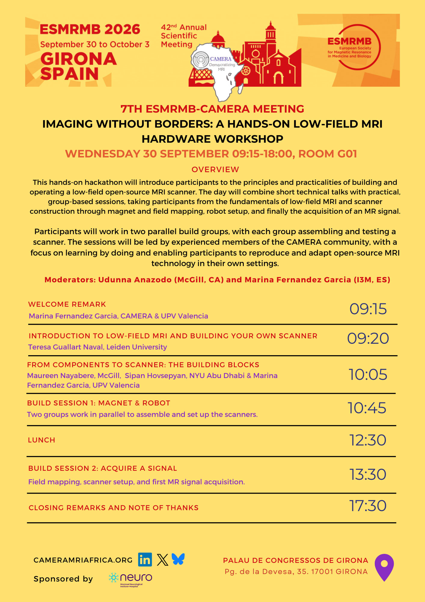
Imaging without Borders: A Hands-On Low-Field MRI Hardware Workshop
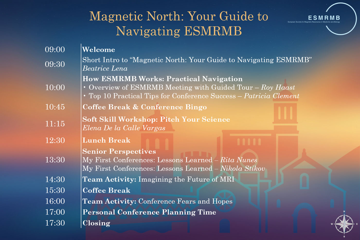
Magnetic North: Your Guide to Navigating ESMRMB
Large international conferences can be overwhelming for newcomers, particularly for those attending alone.
“Magnetic North: Your Guide to Navigating ESMRMB” is a full-day pre-congress event designed to support first-time and early-career attendees by providing a structured and low-pressure introduction to the ESMRMB Annual Meeting. The programme combines practical guidance on navigating the conference with interactive activities aimed at fostering early connections among participants. Sessions include a practical introduction to the structure of the ESMRMB meeting, senior researchers sharing personal conference experiences, guided networking activities, and a soft skill development workshop about pitching science.